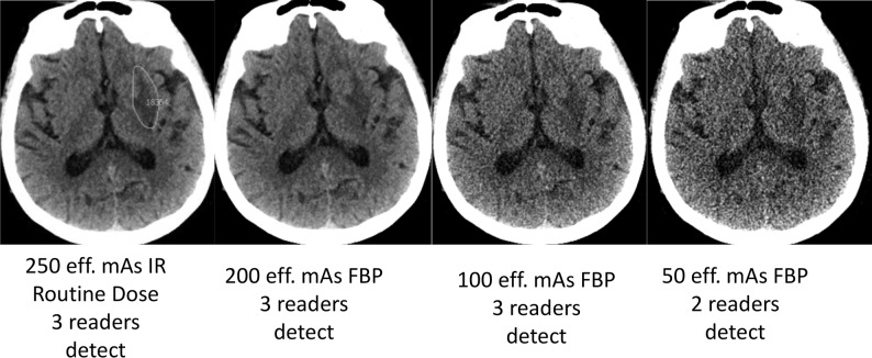
Fig 3.
Acute left lentiform nucleus infarct (green circle indicates reference neuroradiologist markings at routine dose) with corresponding lower-dose FBP CT images along with reader results. The imaging finding on this CT examination evolved with time, with corresponding clinical confirmation of corresponding neurologic deficit by a staff neurologist, and the final diagnosis was recorded as acute left striatal infarct.

