
Figure 3.
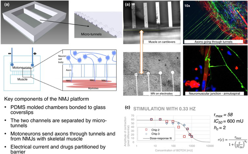
Neuromuscular junction (NMJ) platform. A microtunnel-based system (a) allows the neurons and skeletal muscle to remain in distinct compartments while allowing axons to pass to the muscle side and innervate the myotubes; microfabricated bioMEMS enable direct electrical stimulation of motoneurons and direct measurement of myotube contraction (lower panel). (b) Phase contrast images of myotubes on cantilevers (upper left) and motoneurons on microelectrode array (MEA) electrodes (lower left), immunocytochemistry indicating axons (green) growing through tunnels and forming NMJs with myotubes (red) (right panels). (c) Effect of drugs on the NMJ can be tested in the system, producing a dose–response curve, in this case using BOTOX as the NMJ blocking toxin. As the concentration of BOTOX on the muscle-side increases the amplitude of the myotube contraction decreases. IC50 = 600 mU.