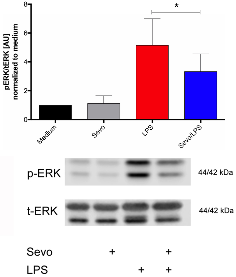
Figure 6.
Expression of extracellular signal-regulated kinase (ERK) phosphorylation (p-ERK) in bone marrow-derived macrophages normalized by total ERK (t-ERK). Cells were stimulated for 8 hours with lipopolysaccharide (LPS) in the presence or absence of 2% sevoflurane, followed by Western blot analysis. Representative Western blots are shown below. Values represent means ± standard deviation; n=7, one-way ANOVA); * p=0.036

