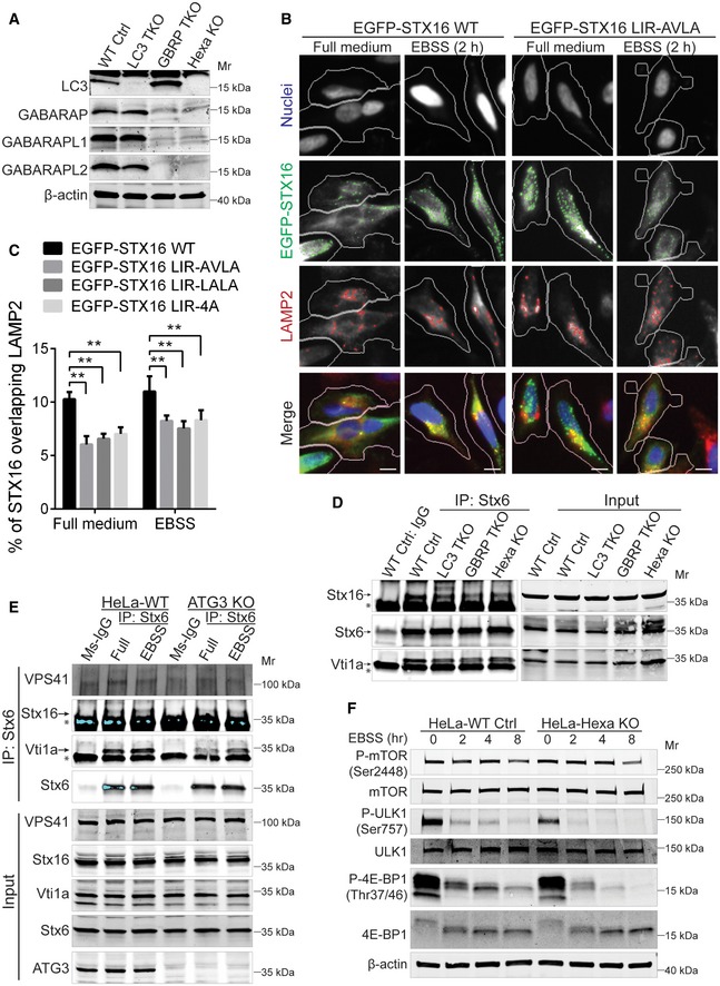
Figure EV5. mAtg8s affect acidification of the endolysosomal system and mTOR activity.

- Western blot analysis of LC3 (LC3A, LC3B, LC3C), GABARAP, GABARAPL1, and GABARAPL2 in WT HeLa or cells knocked out for LC3A, LC3B, LC3C (LC3 TKO), GABARAP, GABARAPL1, and GABARAPL2 (GBRP TKO) or all 6 mATG8s (Hexa KO).
- HeLa cells were transfected with WT or LIR‐mutant EGFP‐tagged Stx16, and subjected to HCM analysis of overlaps between Stx16 and LAMP2. Masks: white, cells transfected with Stx16 identified based on the average intensity of EGFP‐Stx16; green, EGFP‐Stx16 puncta; red, LAMP2 puncta; yellow, overlap between EGFP‐Stx16 and LAMP2. Scale bar: 20 μm.
- HCM quantifications of overlaps between LAMP2 and WT or different types of LIR‐mutant EGFP‐Stx16 transfected into HeLa cells and treated as in (B). Data shown as means ± SEM of percentages of EGFP‐Stx16 overlapping with LAMP2, minimum 200 transfected cells were counted each well from at least 12 wells, 3 independent experiments; **P < 0.01 (one‐way ANOVA).
- Endogenous Co‐IP analysis of the interactions between Stx16, Vti1a, and Stx6 in WT, LC3 TKO, GABARAP TKO, or Hexa KO HeLa cells. * indicates mouse IgG of precipitated mouse anti‐Stx6 antibody.
- Endogenous Co‐IP analysis of the interactions between VPS41, Stx16, Vti1a, and Stx6 in WT, or ATG3 KO cells. * indicates mouse IgG of precipitated mouse anti‐Stx6 antibody.
- WT or Hexa KO HeLa cells were starved in EBSS for indicated time points, and cell lysates were subjected to Western blot analysis of mTOR activity by mTOR substrate phosphorylation.
Source data are available online for this figure.
