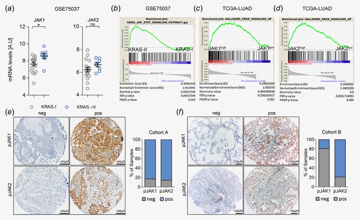
Figure 1.

JAK‐mediated signaling is activated in progressing K‐RAS‐mutated human lung AC. (a) Graph showing relative JAK1 and JAK2 mRNA expression levels in human K‐RAS‐mutated lung AC tissue of stage I (n = 19) versus stage II or higher (≥II, n = 8). Data represent mean ± SEM, A.U (arbitrary units), Student's t‐test, *p < 0.05. (b) GSEA for Kyoto encyclopedia of genes and genomes‐JAK–STAT pathway signature geneset comparing human K‐RAS‐mutated tumors of stage I versus stage II, using tumor versus healthy parenchyma mRNA expression ratios. Data in (a) and (b) were retrieved from the Gene Expression Omnibus (GSE75037). (c) GSEA using gene expression data of the Cancer Genome Atlas‐LUAD cohort and HALLMARK_KRAS_SIGNALING_UP geneset, stratifying patients according to JAK1 and (d) JAK2 expression levels (n = 154). (e) Representative pictures of lung AC biopsies of patients included in cohort A (unknown K‐RAS mutation status; pJAK1 n = 303, pJAK2 n = 318) and (f) cohort B (K‐RAS mutated; pJAK1 n = 26, pJAK2 n = 24) with negative and positive staining reactions for pJAK1 and pJAK2 in tumor cells. Graphs represent percentage of positive and negative cases within the respective cohort. Staining intensities and percentage of positive tumor cells were determined by a board‐certified pathologist (H.P.). [Color figure can be viewed at http://wileyonlinelibrary.com]
