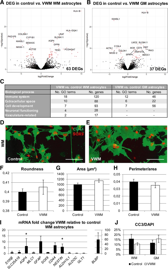
Figure 6.

Vanishing white matter (VWM) human astrocyte subtypes differ in morphology and mRNA expression level from control human astrocyte subtypes. (A, B) Differential expression analysis revealed significant differentially expressed genes (DEGs) between VWM and control in human white matter (WM) astrocytes (A; control, n = 3; VWM, n = 4) and human gray matter (GM) astrocytes (B; control, n = 4; VWM, n = 4). The volcano plots indicate significant DEGs in red, with the 15 most significantly up‐ or downregulated DEGs labeled. (C) Significantly enriched Gene Ontology (GO) terms classified the DEGs in different categories of biological processes. (D, E) Morphology of control (D) and VWM (E) human WM astrocytes was visualized using CD44 and SOX9 immunostaining. (F–H) Morphological analysis showed differences in roundness (F), area (G), and perimeter corrected for area (H) between VWM and control human WM astrocytes based on the CD44 immunostaining. (I) A quantitative polymerase chain reaction on VWM human WM astrocytes (n = 4) showed differential expression of S100B, SLC25A18, MLC1, SOX9, CD44, NESTIN, ALDH1L1, ALDOC, SLC1A2, AQP4, GFAP, and BLBP relative to control human WM astrocytes (n = 4). *p < 0.05. (J) The cleaved caspase 3 (CC3)‐positive percentage of the 4,6‐diamidino‐2‐phenylindole (DAPI)‐positive cells was determined in oligodendrocyte cultures containing media conditioned by GM or WM astrocytes derived from control (n = 4) and VWM (n = 4) lines. Bars represent mean ± standard error of the mean.
