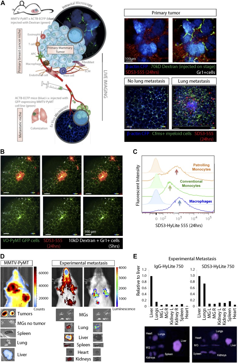
Figure 3. SDS3 concentrated within and around primary tumor and metastatic foci.
(A) Left: schematic of experimental setup for imaging MMTV-PyMT; ACTB-ECFP i.v. injected with VO-PyMT-GFP-Luc cells and SDS3-HyLite 750. Right: top—primary tumor confocal microscopy 24 h after SDS3 injection into MMTV-PyMT; ACTB-ECFP mice shows SDS3 present within tumor stroma (n = 8). See Video 1 for full video of still shots shown; bottom—metastatic lung confocal microscopy 24 h after SDS3 injection into MMTV-PyMT; ACTB-ECFP mice shows SDS3 accumulating at the metastatic sites as compared with lung parenchyma (n = 5). See Video 2 for full video of still shots shown. (B) An MMTV-PyMT; ACTB-ECFP mouse was i.v. injected with VO-PyMT-GFP-Luc cells. 1 wk later, SDS3-HyLite 555 was injected 24 h before imaging followed by near-infrared (NIR) 10-kD dextran and anti-Gr1 antibody 5 h before imaging. Representative images of confocal microscopy show NIR 10-kD dextran and anti-Gr1 antibody (white) accumulate around VO-PyMT metastasis (green) and SDS3-HyLite 555 (red) (n = 5). See Videos 3 and 4 for full video of still shots shown. (C) A representative flow cytometry analysis of the lungs of MMTV-PyMT mice 24 h after i.v. injection of VO-PyMT-GFP-Luc cells and treated with SDS3 or IgG isotype control. (D) Left: 1 × 10⁵ VO-PyMT-GFP-Luc cells were i.v. injected into MMTV-PyMT mice along with SDS3-HyLite 750. MMTV-PyMT mice IVIS image depicts high intensity of signal resting within the lungs 24 h after injection (n = 3). Right: 1 × 10⁶ VO-PyMT-GFP-Luc cells were i.v. injected into WT mice along with SDS3-HyLite 750. Fluorescent IVIS whole-body imaging shows localization of SDS3-HyLite 750 and bioluminescent IVIS whole-body imaging shows VO-PyMT-GFP-Luc cells seeding within the lungs of WT mice (n = 3). (E) Ex vivo fluorescent and bioluminescent imaging of various organs from WT mice i.v. injected with 1 × 10⁶ VO-PyMT-GFP-Luc shows that the strongest signal was detected in the lung, indicating SDS3 accumulation at the metastatic site with no retention of IgG isotype control within the lungs (n = 3 IgG-HyLite 750, n = 3 SDS3-HyLite 750).

