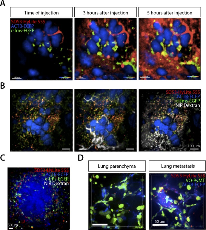
Figure S3. SDS3 accumulation around metastatic foci.
(A) Intravital confocal microscopy of the primary tumor in MMTV-PyMT; ACTB-ECFP mouse i.v. injected with 1 × 10⁵ VO-PyMT-GFP-Luc cells and SDS3-HyLite 555. SDS3-HyLite 555 (red) is seen to leak from the tumor vasculature and accumulate in the stroma (n = 8). See Video 7 for full video of still shots shown. (B) Intravital confocal microscopy of the primary tumor in MMTV-PyMT; ACTB-ECFP mouse 2 wk after i.v. injection of 1 × 10⁵ VO-PyMT-GFP-Luc cells. SDS3-HyLite 555 was injected 24 h before imaging followed by NIR 10-kD dextran and anti-Gr1 antibody 5 h before imaging. Images show NIR 10-kD dextran (white) and anti-Gr1 antibody (green) accumulation around tumor stroma (blue), and SDS3-HyLite 555 (red) also accumulated around tumor stroma and vasculature (n = 8). See Video 8 for full video of still shots shown. (C) Intravital confocal microscopy of the lungs in MMTV-PyMT; ACTB-ECFP mouse 2 wk after i.v. injection of 1 × 10⁵ VO-PyMT-GFP-Luc cells. SDS3-HyLite 555 was injected 24 h before imaging followed by NIR 10-kD dextran and anti-Gr1 antibody 5 h before imaging. Images show NIR 10-kD dextran (white) and anti-Gr1 antibody (green) accumulation around VO-PyMT metastasis (blue), and SDS3-HyLite 555 (red) also accumulates around VO-PyMT metastasis (n = 5). (D) Intravital confocal microscopy of the lungs in MMTV-PyMT; ACTB-ECFP mouse 2 wk after i.v. injection of 1 × 10⁵ VO-PyMT-GFP-Luc cells. SDS3-HyLite 555 (red) accumulates around VO-PyMT metastasis (green) (n = 5).

