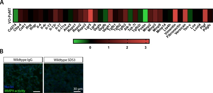
Figure S4. VO-PyMT cell gene expression profile.
(A) qPCR analysis of VO-PyMT cells relative to EpH4, a normal mammary epithelial cell line. VO-PyMT tumor cells had elevated levels of various pro-metastatic factors that enhance metastatic dissemination and colonization such as Mmp2/3/9 alongside Vimentin and Versican, whereas showing decreased levels of cytokines associated with immune recruitment such as Cd274, Ifnγ, and Ifnγr. (B) Representative images of in situ zymography in experimental metastasis of IgG- and SDS3-treated WT lungs at 9-wk-old after a 3-wk chase of i.v. injected VO-PyMT cells. Cell nuclei are stained with DAPI (blue) and MMP2/9 activity shown in green.

