Figure 3.

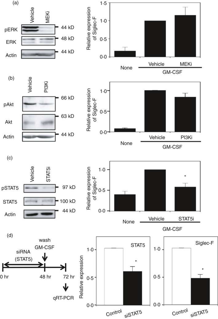
Enhancement of Siglec‐F expression by granulocyte–macrophage colony‐stimulating factor (GM‐CSF) depends on the signal transducer and activator of transcription 5 (STAT5) pathway. (a) Effects of the mitogen‐activated protein kinase kinase (MEK) inhibitor (PD0325901). (Left) Confirmation of MEK inhibition by a reduction in the phosphorylation of extracellular signal‐regulated kinase (ERK). Macrophage colony‐stimulating factor‐differentiated bone‐marrow‐derived macrophages (M‐BMDMs) were stimulated with GM‐CSF for 30 min. A representative result of two independent experiments is shown. (Right) The lack of inhibition of Siglec‐F expression by the MEK inhibitor. Siglec‐F expression was measured by qRT‐PCR after a 24‐hr stimulation. The vehicle control [dimethyl sulfoxide (DMSO) 0·1%] was regarded as 1. Data are the mean ± SE of three independent experiments. (b) Effects of the phosphoinositide 3‐kinase (PI3K) inhibitor (wortmannin). (Left) Confirmation of PI3K inhibition by a reduction in the phosphorylation of Akt. Cells were stimulated with GM‐CSF for 30 min. A representative result of three independent experiments is shown. (Right) The lack of inhibition of Siglec‐F expression by the PI3K inhibitor. Siglec‐F expression was measured by qRT‐PCR after a 24‐hr stimulation. The vehicle control (DMSO 0·1%) was regarded as 1. Data are the mean ± SE of five independent experiments. (c) Effects of the STAT5 inhibitor. (Left) Confirmation of STAT5 inhibition by a reduction in phosphorylation. Cells were stimulated with GM‐CSF for 30 min. A representative result of two independent experiments is shown. (Right) The inhibition of Siglec‐F expression by the STAT5 inhibitor. Siglec‐F expression was measured by qRT‐PCR after an 8‐hr stimulation. The vehicle control (DMSO 0·5%) was regarded as 1. Data are the mean ± SE of three independent experiments. *P < 0·05 versus the vehicle by Student's t‐test. (d) Enhancing effects of GM‐CSF on Siglec‐F expression are inhibited by the knockdown of STAT5. (Left) Schematic presentation of the experiment. M‐BMDMs were transfected with STAT5 or control small interfering RNA (siRNA). Cells were washed after a 48‐hr culture and stimulated with GM‐CSF for an additional 24 hr. The expression of STAT5 and Siglec‐F was measured by qRT‐PCR. (Middle) Knockdown efficiency of STAT5. (Right) Inhibition of Siglec‐F expression by STAT5 siRNA. The mRNA level in cells transfected with control siRNA was regarded as 1. Data are the mean ± SE of three independent experiments. *P < 0·05 versus control siRNA by Student's t‐test.
