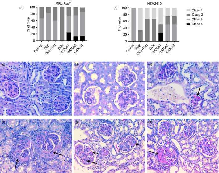
Figure 8.

Histopathology of kidneys. The extension of glomerular, tubule interstitial and vascular injury was evaluated using the current classification for patients with lupus nephritis. The score for grading active lesions was a modification of the activity index used currently in renal diagnostic pathology. Grade of glomerulonephritis according to International Society of Nephrology/Renal Pathology Society 2003 classification of glomerulonephritis is shown for each animal group for (a) MRL‐Faslpr mice and (b) NZM2410 mice (for three or four animals per group depending on the case). Representative images for each class observed are shown at 40× with Periodic acid–Schiff stain (c–h). (c) Class I/Minimal mesangial lupus nephritis. High view with normal glomeruli by light microscopy. (d) Class II/Mesangial proliferative lupus nephritis. Light microscopy high view showing a slightly mesangial hypercellularity with focal mesangial matrix expansion. (e) Class III A/Active lesions: Focal (lesion involving < 50% of glomeruli) proliferative lupus nephritis. Arrow shows intraluminal immune aggregates (hyaline thrombi). (f) Class III (A/C)/Active and chronic lesions: Focal proliferative and sclerosing lupus nephritis. Arrows indicate fibrocellular crescents. (g) Class IV‐S (A/C)/Active and chronic lesions: Diffuse (lesion involving ≥ 50% glomeruli) segmental proliferative and sclerosing lupus nephritis. Active lesions are karyorrhexis (long arrow) and subendothelial deposits identifiable by light microscopy (wireloops, short arrow). (h) Class IV‐S (A/C)/Active and chronic lesions: Diffuse segmental proliferative and sclerosing lupus nephritis. Arrow shows segmental glomerular sclerosis.
