Figure 1.
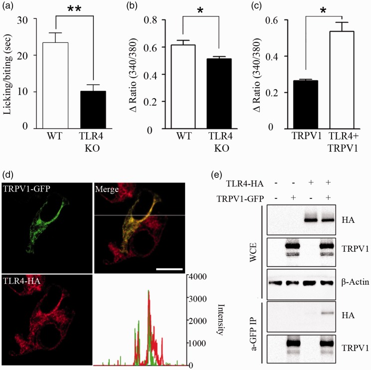
Direct interaction between TLR4 and TRPV1. (a) To confirm the effect of TLR4 expression on TRPV1-activated mice behavior, WT and TLR4 KO mice were administered capsaicin (1.6 µg) by intraplantar injection on the right hind paw. Licking/biting time of the right hind paw was measured for 5 min after capsaicin injection (n = 7, **p < 0.01). (b) DRG neurons from WT and TLR4 KO mice were cultured and treated with capsaicin (1 µM), and intracellular calcium was monitored by calcium imaging assays (n = 5, *p < 0.05). (c) HEK293T cells transiently overexpressed with TRPV1-GFP or TRPV1-GFP plus TLR4-HA (n = 6) were loaded with Fura-2 AM. Cells were treated with capsaicin (10 µM), and intracellular calcium level was measured by spectrofluorophotometer population assay. Mean with SEM are shown (n = 6, *p < 0.05). (d) TRPV1 and TLR4 were transiently overexpressed in HEK293T cells, and sub-cellular fluorescence intensities were analyzed under a confocal microscope. The fluorescence intensities of TRPV1 and TLR4 along the x-axis are shown in a graph (right). The subcellular localization of TRPV1 merged with that of TLR4. Scale bar, 20 µm. (e) HEK293T cells were transfected with TLR4-HA, TRPV1-GFP, or TLR4-HA plus TRPV1-GFP expression vectors. Total cell extracts were immunoprecipitated with anti-GFP antibody, and then TLR4 expression was measured using anti-HA antibody (lower two panels). In addition, expression levels of TLR4, TRPV1, and β-actin in the WCE were measured (upper three panels). Representative gel pictures are shown (n = 3).
WT: wild-type; TLR4: toll-like receptor 4; KO: knockout; TRPV1: transient receptor potential V1; GFP: green fluorescent protein; WCE: whole-cell extracts.

