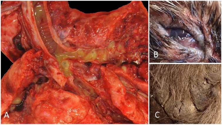
Figure 2.
Gross anatomic lesions of a distinct clade of canine distemper virus (CDV) infection in wildlife from New Hampshire and Vermont. A. CDV in fisher A. The terminal trachea and mainstem bronchi are opened along with the surrounding parenchyma, revealing severe, multifocal-to-coalescing areas of consolidation, and associated and surrounding erythema, with mucopurulent exudate in the airways. B. Chronic, diffuse, mucoid and proliferative conjunctivitis and palpebritis in a raccoon. C. Hyperkeratosis and degeneration of the footpads in fisher C, characterized by an irregular and pronounced surface that has multiple irregular fissures, a.k.a. “hardpad.”

