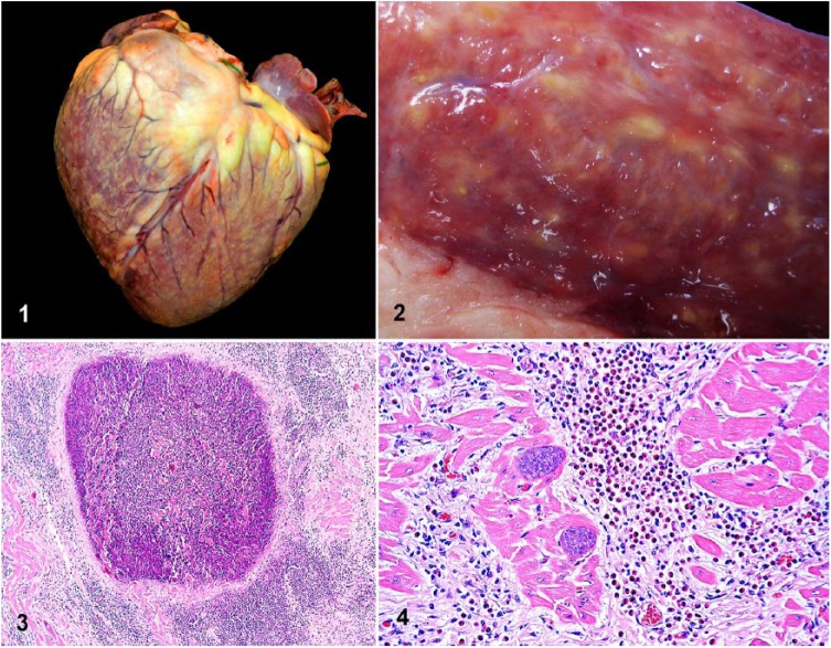
Figures 1–4.
Eosinophilic granulomatous myocarditis in a heifer. Figure 1. Myriad 1–3-mm, irregular, yellow-green coalescing foci disseminated throughout the myocardium are visible from the epicardial surface. Figure 2. Transmural section of the left ventricular free wall with granulomas and white streaks throughout the myocardium. Figure 3. A myocardial granuloma composed of a well-demarcated central area of necrosis surrounded by dense interstitial mixed inflammatory cell infiltrates and fibrosis. H&E. Figure 4. Abundant eosinophils, fewer lymphocytes, and macrophages infiltrate the myocardial interstitium, which is also expanded by fibrosis. Adjacent cardiomyocytes contain intact intrasarcoplasmic, thin-walled, basophilic Sarcocystis cruzi cysts. H&E.

