Figures 1–4.
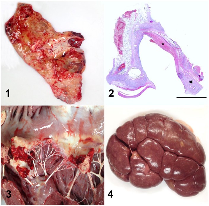
Cranial superficial epigastric vein (CSEV) phlebitis and septicemia in dairy cows. Figure 1. The intimal surface of the CSEV (arrows) of case 4 is roughened and covered by thin layers of fibrin (asterisk). The adjacent areas were markedly swollen and firm, as a result of edema and fibrosis. Figure 2. The wall of the CSEV of case 4 was markedly thickened by fibrosis and moderate numbers of lymphocytes, plasma cells, and macrophages, mainly in the tunica adventitia (arrowhead). There were extensive areas of endothelial loss, covered by strands of fibrin, and expansion of the subendothelial spaces with fibrin, neutrophils, and cell debris (asterisk). H&E. Bar = 5 mm. Figure 3. Vegetative endocarditis of the mitral valve of case 4. Figure 4. The surface of the renal cortex of case 4 contained irregular pale tan areas (infarcts) and yellow, 0.1–0.3-mm diameter nodules of embolic nephritis.

