Figure 7.
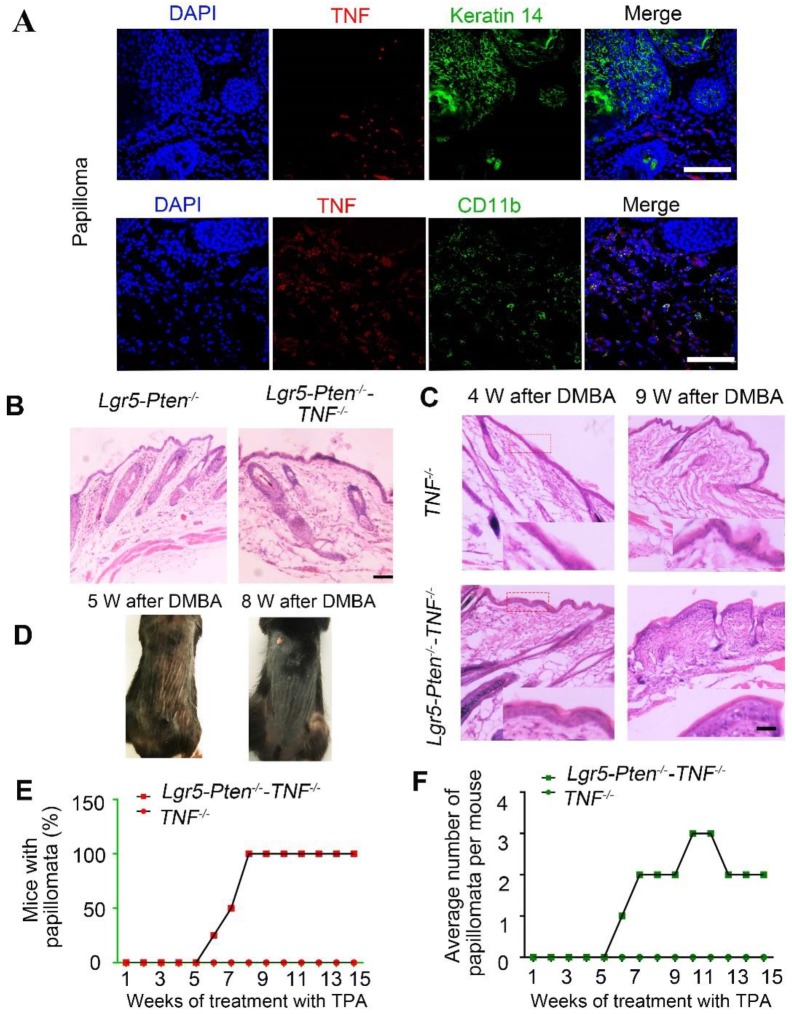
TNF is necessary for Pten loss induce tumor formation. (A) TNF expression was detected in papillomata and was largely present in CD11b+ cells. (B) 40 days after injection tamoxifen for three times, histological analysis (HE staining) of the dorsal skin of Lgr5-Pten-/-mice and Lgr5-Pten-/--TNF-/-mice showed HF hyperplasia (telogen phase). (C) 4 and 9 weeks after DMBA/TPA treatment, epidermal hyperplasia in the dorsal skin of Lgr5-Pten-/--TNF-/- mice was more obvious than in TNF-/-mice. (D) Representative images showing papillomata in Lgr5-Pten-/--TNF-/- mice 5 weeks and 8 weeks after DMBA/TPA treatment. (E) The incidence of skin tumor development in TNF-/- mice and in Lgr5-Pten-/--TNF-/- mice. (E) The average number of papillomata per mouse in TNF-/- mice (n=8) and in Lgr5-Pten-/--TNF-/-mice (n=8). Scale bars, 100 μm. W, weeks.

