Figure 1.

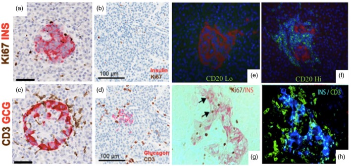
Heterogeneous islet morphology and immune cell infiltration across disease duration and according to age of T1D onset. Representative islets were imaged from serial paraffin sections stained for Ki67 plus insulin (INS) and CD3 plus glucagon (GCG) for a 13‐year‐old donor at T1D onset (nPOD 6228; a & c) and a 6 year old with 3 years T1D duration (nPOD 69; b & d) 47, 48. CD3+ infiltrate is present in both donors (c, d), though insulin containing islets are only observed in the first donor (a, b). Scale bars: a and c, 50μm; b and d, 100μm 47, 48. Pancreas samples from donors with recent‐onset T1D stained for CD20 (green) and glucagon (red), and nuclei (DAPI) exhibit differences in infiltrate composition, which can separate subjects based on hyper‐immune CD20Hi (nPOD 6052; e) and pauci‐immune CD20Lo profiles (nPOD 6070; f) 50. Histology of a 46 year old donor with ≥3 islet AAb shows both Ins+Ki67– β‐cells and Ins+Ki67+ cells replicating β‐cells (g, arrows) within islets that contain CD3+ T cell infiltrate (h) 51. Figures have been reprinted with permission from the American Diabetes Association 47, 48, 50, 51.
