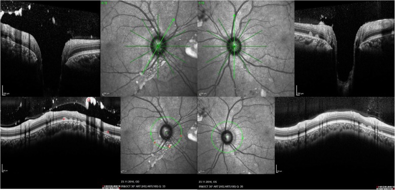
Fig. 6.
OCT of posterior segment abnormalities in another Gaucher patient. Optical coherence tomography of the right and left eye showing atrophy (←) and subretinal deposits (*) from 2 to 8 o’clock in the peripapillary area of the right eye. The left eye is less affected. Both eyes show remarkable vitreous opacities (o). Quality indices: right eye 31/33, left eye 32/26

