Fig. 1.
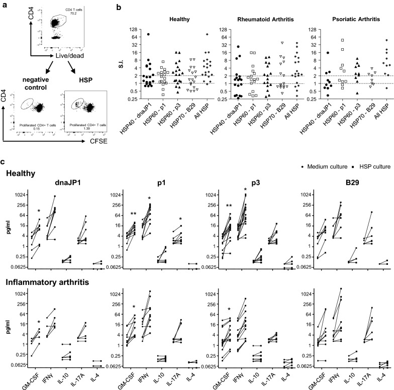
Inflammatory arthritis (IA) patients have pro-inflammatory HSP-specific CD4+ T-cells. Cell proliferation dye (CFSE/CTV)-labelled PBMC of healthy controls and IA patients were cultured with pan-DR-binding HSP peptides: DnaJP1, HSP60p1, HSP60p2 and B29 for 9 days. a, b Percentage of CFSE/CTV-negative live CD4+ T-cells was measured using flow cytometry. Gating example (a) and graphs with stimulation index (SI) b are shown. SI was measured by dividing the percentage of CFSE/CTV− CD4+ T-cells of HSP culture by the percentage of CFSE/CTV− CD4+ T-cells medium control (i.e. no peptide added) culture. SI > 2 was considered as increased above background proliferation. ‘All HSP’ indicates best HSP response per donor. c Cytokine secretion in culture supernatants was measured using MSD immunoassay. Left circle indicates cytokine concentration in medium control culture, right square indicates cytokines concentration in HSP culture. Two-tailed paired Student T-test was used. *p ≤ 0.05, **p ≤ 0.01

