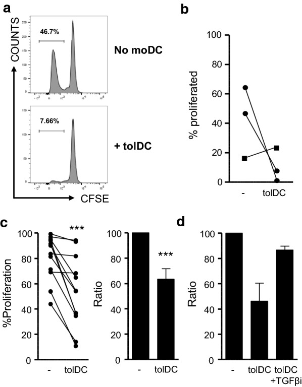
Fig. 5.

tolDC inhibit bystander NK-cell proliferation. Cell proliferation dye (CFSE/CTV)-labelled PBMC of IA patients were cultured with an HSP-peptide pool (HSP60p1, HSP60p2 and B29; 4 μg/ml per peptide) (a, b) or CA (1:1000) (c, d) and tolDC (tolDC) or without moDC (−) for 9 days. a–d Percentages of CFSE/CTV− life NK-cells were measured using flow cytometry. Gating strategy (a) HSP-peptide graph (b) and CA graphs (c, d) are shown. (d) TGF-βRI (ALK5) inhibitor (SB-505124) was added at 1 µM. (N = 2). Ratios were measured by dividing the percentage of CFSE/CTV− NK-cells of tolDC cultures by the percentage of CFSE/CTV− NK-cells of non-moDC cultures. Two-tailed paired Student T-test was used. ***p ≤ 0.001
