
Fig. 3.
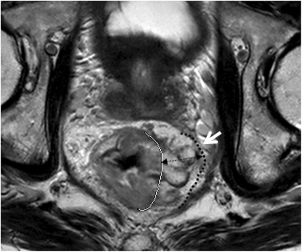
Oblique axial T2-weighted imaging of a rectal mucinous adenocarcinoma (intermediate to high signal intensity) presenting as bulky mass showed significant tumor infiltration beyond the muscularis propria; the maximal extramural depth (MEMD, double-headed arrow) was over 10 mm. Meanwhile, the invasive border of rectal mass bordering the mesorectal fascia (white arrow) which leaded to a CRM of 0 mm. White line = muscularis propria border. Black dashed line = the mesorectal fascia