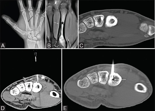
Figure 1 (A-E).

(A) AP radiograph showing periosteal reaction on the medial side of the 2nd metacarpal. (B) Coronal PD fat saturated MRI of the hand showing periosteal reaction on the ulnar side of the 2nd metacarpal (white arrow) with marrow edema (black star). (C) Axial non contrast CT image of the hand demonstrating the nidus (white arrow). (D) Axial CT of the hand with a 20 G needle (white arrow), placed on the dorsal surface of the metacarpal for infusion of D5W while the ablation is in progress to prevent skin burns. The infused fluid is seen creating a buffer between the skin and the bone (white star). (E) Axial CT demonstrating the RF probe in the OO nidus (white arrow)
