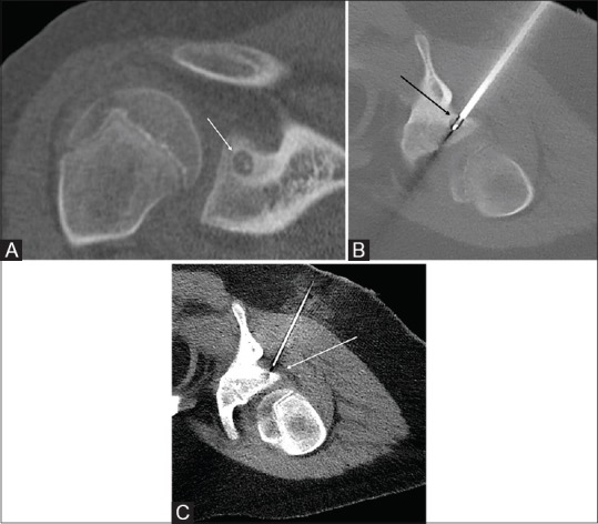
Figure 2 (A-C).

(A) CT coronal reconstruction shows a right glenoid osteoid osteoma (white arrow). (B) Intra-procedural CT showing the ablation probe with tip within the nidus (black arrow). (C) Intraprocedural CT with a 20 G spinal needle placed in the spino-glenoid notch (white arrow) to facilitate D5W injection during the ablation for protection of the suprascapular nerve
