
Figure 1 (A and B).
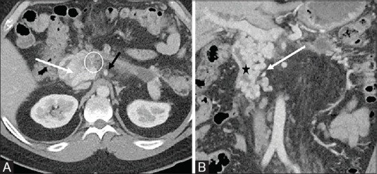
Axial (A), coronal (B) contrast enhanced abdominal CT showing large duodenal varices (white arrows). Severe desmoplastic reaction in the region of the superior mesenteric vein, causing complete occlusion (white circle). Superior mesenteric artery patent (black arrow). The black star corresponds to the duodenal lumen