Figure 4 (A and B).

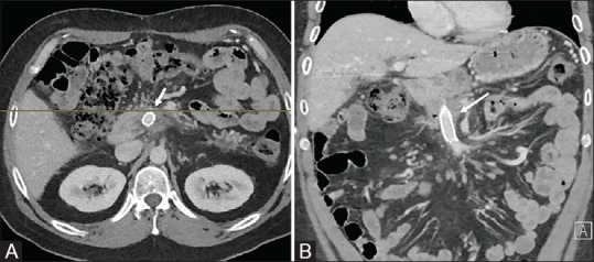
Contrast enhanced axial (A) and coronal (B) CT images obtained 3 months post stent placement. Patent SMV stent (arrow). No varices seen

Contrast enhanced axial (A) and coronal (B) CT images obtained 3 months post stent placement. Patent SMV stent (arrow). No varices seen