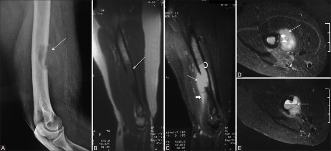
Figure 1 (A-E).
A 50-year-old female with swelling in the right forearm (Case 1). (A) Lateral radiograph of arm including elbow showing expansile, eccentric, cortical-based lytic lesion with thin shell of cortex in lower diaphysis of right humerus (arrow) B-D: Sagittal T1W (B), T2W fat saturated (C), Axial T2W fat saturated (D and E) MR images is showing lesion in lower end of humerus which is hypointense on T1W (arrow, B), hyperintense on T2W (arrow, C) associated with a soft tissue component (D) having blood-fluid levels (arrow, E) with extensive marrow (curved arrow, C) and soft tissue edema (block arrow, C)

