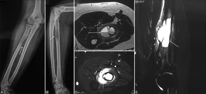
Figure 5 (A-E).
A 16-year-old young female with swelling in the right forearm. (A and B) AP and lateral radiographs of the forearm showing expansile, eccentric cortical based lytic lesion with a thin-shelled cortex arising from the proximal diaphysis of radius (arrow, A and B). (C-E) Axial T2W, Axial T2W fat-saturated and Sagittal T2W fat-saturated MR images showing large exophytic soft tissue component with blood-fluid levels (arrow, C) with extensive soft tissue (arrow, D) and marrow oedema (arrow, E)

