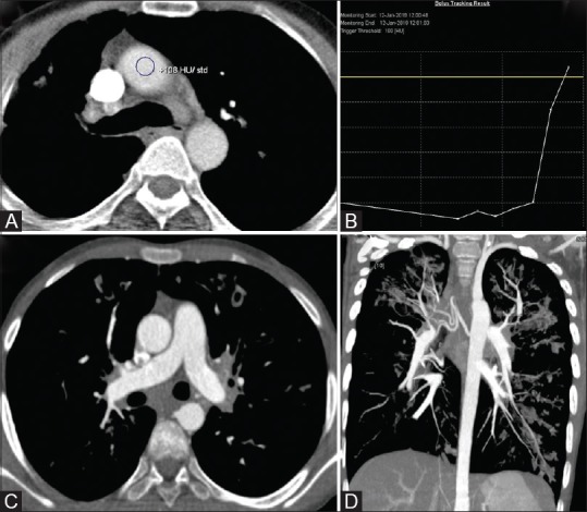
Figure 12 (A-D).

Single phase split-bolus DECT angiography in a case of hemoptysis. (A and B) ROI positioning in ascending aorta with trigger at 100HU. (C and D) Simultaneous optimal opacification of aorta, pulmonary artery and their branches is seen
