
Figure 14 (A-E).
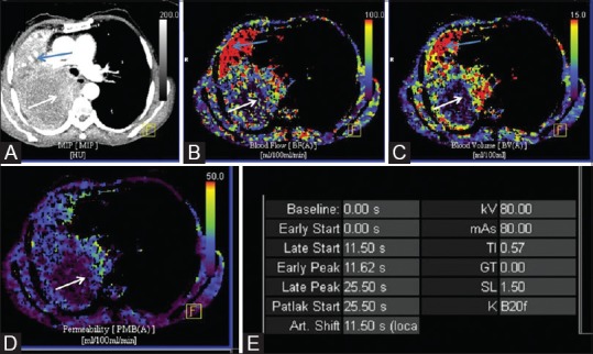
Dynamic perfusion CT of large necrotic mass to choose appropriate site of biopsy. (A) Axial maximum intensity projection (MIP) image in mediastinal window shows large necrotic mass in right hemithorax, with peripheral thick irregular nodular enhancement (white arrow) and associate collapse of lung (blue arrow). Color-coded blood flow (B), blood volume (C) and permeability (D) maps showing increased perfusion in peripheral nodular areas of enhancement (white arrow) with hypoperfused necrotic centre. Distally collapsed lung shows homogeneous high perfusion (blue arrow). (E) Image showing technical parameters of the acquisition