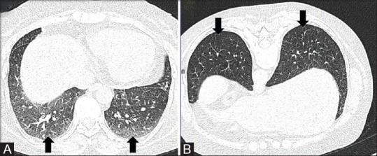
Figure 4 (A and B).

NCCT variants. NCCT chest in supine (A) position shows ground glass opacities in posterior basal segments of bilateral lower lobes (arrows). CT performed in prone position (B) shows clearing of the GGOs suggestive of dependent densities (arrows)
