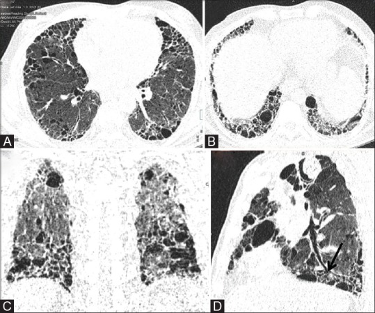
Figure 6 (A-D).

LDCT for evaluation of ILD. (A) Axial image of LDCT in high resolution reconstruction algorithm in a 65-year old chronic smoker with suspected ILD. (B and C) Routine axial and coronal reconstruction in lung window allow evaluation of cranio-caudal and axial distribution of reticulations, interlobular septal thickening and macrocysts. (D) Curved MPR along the axis of bronchus (black arrow) allows distinction between honeycombing or macrocysts and traction bronchiolectasis
