Figure 8 (A-C).

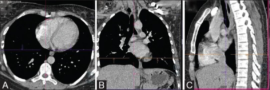
(A-C) Contrast-enhanced CT chest (Routine). Axial, coronal and sagittal reformations in standard mediastinal window. The cross-bar allows three-dimensional localization of lesion

(A-C) Contrast-enhanced CT chest (Routine). Axial, coronal and sagittal reformations in standard mediastinal window. The cross-bar allows three-dimensional localization of lesion