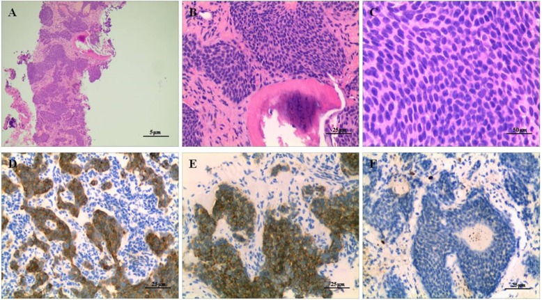
Fig. 4.
Histopathological examination of the needle biopsy (H&E staining) of the 2nd rib shows the tumor consisting of cellular nests of small round cells, no mitoses, original magnification, × 40 (a), × 200 (b), × 400 (c). These cells were positive for synaptophysin, original magnification, × 200 (d), CD56, × 200 (e) and Ki-67 was less 1% upon immunostaining, × 200 (f)

