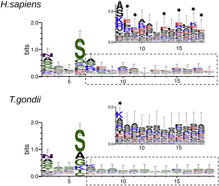
Figure 6. Web logo of HsNMT and TgNMT peptides substrates.
Amino acid alignment of HsNMT and TgNMT peptide substrates from positions 3 to 18. Residues are highlighted according to their chemistry (polar: green; neutral: purple; basic: blue; acidic: red; hydrophobic: black). For both enzymes, a zoomed view over positions 6 to 18 (dotted lines) is shown; residues are highlighted by charge (neutral: black; positive: blue; negative: red). Stars indicate differences.

