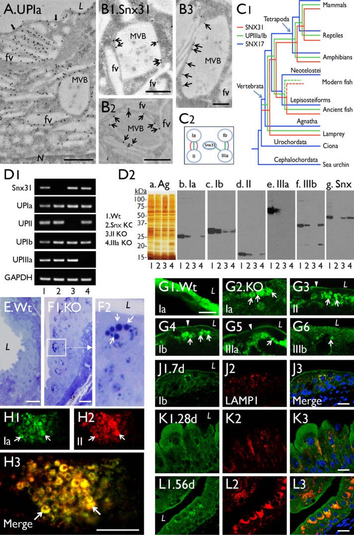
FIGURE 1:
Snx31 knockout leads to the loss of MVBs, reduced uroplakin content, and accumulation of lysosomes (related to Supplemental Figure 1). (A) Immunogold-EM localization of a mouse umbrella cell showing the association of uroplakins with apical plaques (black arrows), fusiform vesicles (FVs), and multivesicular body (MVB)-associated plaques. (B1–B3) Immunogold-EM localization of Snx31 (arrows) showing its association with the plaques of the MVB, but not with FVs. (C1) Coappearance of uroplakin and Snx31 genes during vertebrate evolution, with Snx17 gene as a “control” (see Materials and Methods). (C2) Evolutionary congruence analyses showing the sequence coevolution between genes encoding uroplakins Ia/II, Ib/IIIa, and Snx31-UPIIIa (the three colored connecting lines indicate coevolution as detected by different programs; see Materials and Methods). (D1) Detection of the Snx31, UPIa, II, Ib, IIIa, and Gapdh mRNA by RT-PCR in (1) wild type, (2) Snx31-KO, (3) UPII-KO, and (4) UPIIIa-KO. (D2) Immunoblot analyses of total membrane proteins (samples of lanes 1–4 corresponding to those of D1) after staining using (a) silver nitrate (Ag) or (b–g) monospecific antibodies as indicated. Five mice per line were sacrificed for protein collection. (E, F) Toluidine blue staining of Wt (E) and Snx31-KO (F1) urothelial sections showing the accumulation of cytoplasmic droplets (arrows) in the latter (F2 is a magnified view of the boxed area in F1). (G) Immunofluorescence staining of Wt (G1) and Snx31-KO urothelial sections (G2–G6) for uroplakins Ia, II, Ib, IIa, and IIIb as indicated; note in the KO urothelium the decrease in apical and (cytoplasmic) vesicle-associated uroplakins, which are now concentrated in droplet-like structures (arrows). (H) Double staining of UPIa and UPII in a Snx31-KO urothelial section showing their precise colocalization (arrows). (J–L) Double staining of UPIb (green) and lysosomal marker LAMP1 in urothelia of Snx31-KO mice that are 7, 28, and 56 d old, respectively. Note the progressively increasing accumulation of uroplakin-containing droplets surrounded by lysosomes. Scale bars = 0.5 µm in A and B; 10 µm in G and H; and 20 µm in E, F, J, K, and L.

