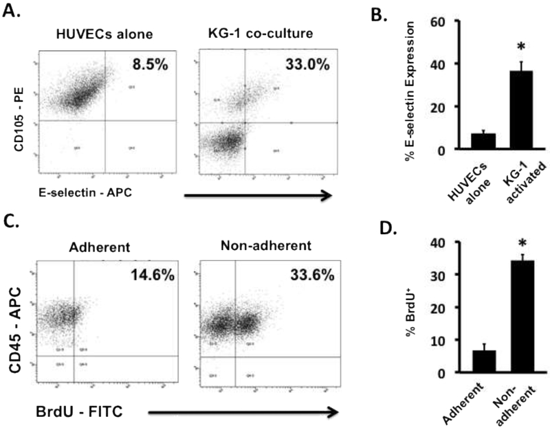
Figure 1. Leukemia cells activate resting ECs resulting in altered leukemia cell proliferation.
(A) Representative flow cytometry plots showing E-selectin levels on KG-1 activated HUVECs and non-activated HUVEC controls. (B) The levels of E-selectin expression on the surface of ECs showed significant increases when activated with KG-1 cells in co-culture. * p < 0.05 (C) Representative flow cytometry plots showing BrdU uptake by adherent and non-adherent AML cells in contact co-cultures of HUVECs and KG-1 cells. (D) BrdU uptake in non-adherent KG-1 cell populations was significantly higher in comparison to adherent populations indicating a proliferative phenotype. * p < 0.05

