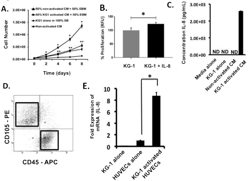
Figure 2. Leukemia activated ECs secrete IL-8 which enhances leukemia cell expansion.
(A) Growth curves of KG-1 cells grown in different media are shown. Supplementing base EBM media with 50% (by volume) activated CM induces significant levels of KG-1 cell growth in comparison to all other cultures tested including those supplemented with non-activated CM. Cells from each culture cohort were enumerated every 2-days over an 8-day culture period. * p < 0.05 versus all other cultures; ** p < 0.05 versus 100% EBM cultures. (B) KG-1 proliferation was significantly enhanced when media was supplemented with IL-8. (C) Fresh, unfrozen supernatants from non-activated and activated co-cultures were evaluated for the production of IL-8 by ELISA. Higher IL-8 concentrations were observed in activated CM. As controls, supernatants from KG-1 alone cultures and pure EGM-2 media were analyzed. Values were extrapolated from standard curves with linear detection limits of 10–3300 pg/mL. ND indicates non-detectable levels. (D) Flow cytometry-based sorting was used to isolate ECs from activating co-cultures. Flow cytometry plots identify gates established for sorting. Representative plots are shown. ECs were isolated based on CD105 (PE) expression while KG-1 cells were identified using CD45 (APC). (E) Sorted ECs were analyzed for mRNA expression levels for IL-8 using qRT-PCR. Non-activated ECs and KG-1 cells alone were analyzed as negative controls. * p < 0.05 versus ECs alone

