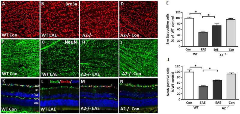
Figure 3: A2 deletion protects against EAE-induced RGC loss.
Representative confocal images showing the immunolabeling of retinal flat mounts with Brn3a (A-D) and NeuN in the GCL layer (F-I). Quantitative analysis demonstrating significant loss of NeuN-positive (E) and Brn3a positive (J) cells in the GCL in response to EAE induction. A2 deletion significantly protected against the EAE induced neuronal loss. Data are presented as mean ± SEM. * (P < 0.01); # (p< 0.05). N = 6-7 per group. Scale bar: 50 μm. K-N) Confocal images of retinal cryostat sections immunostained with NeuN, Brn3a and DAPI. Arrows indicate areas of cell loss. Scale bar: 50 μm. GCL, ganglion cell layer; INL, inner nuclear layer; IPL, inner plexiform layer; ONL, outer nuclear layer; OPL, outer plexiform layer. N= 4-6, and representative images are presented.

