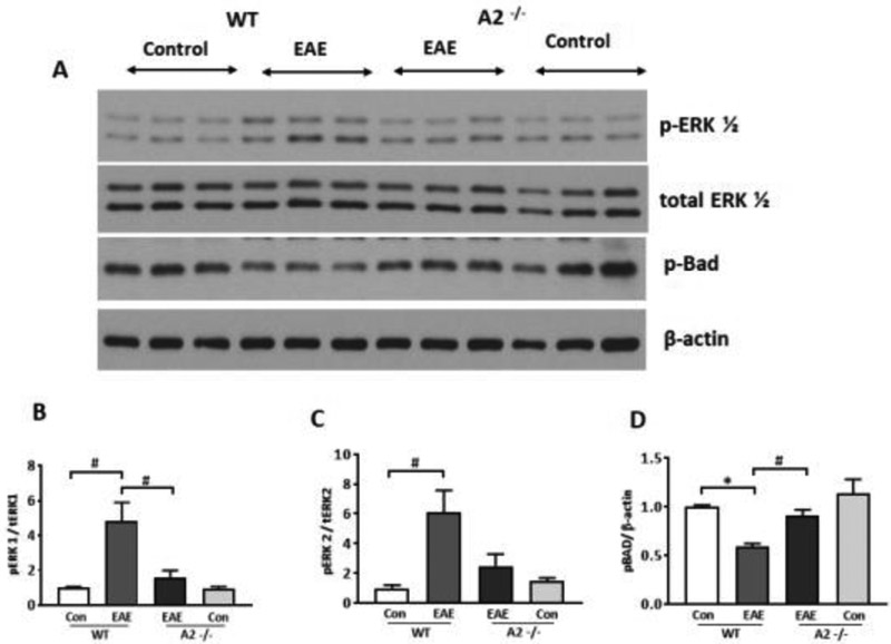
Figure 9: Deletion of A2 reduced EAE induced stress signaling in the retina.
A) Western blot studies showing increased levels of p-ERK1/2 and reduced p-BAD levels in WT EAE retina. These changes were reversed in A2−/− EAE retina. B-D) Quantitative analysis of western blots demonstrate significantly increased levels of p-ERK1/2 and reduced levels of p-BAD in the WT EAE retina compared to WT control. Deletion of A2 altered the EAE-induced increase in p-ERK1 levels and reduction in p-BAD levels. Data are presented as mean ± SEM. *P < 0.01; #p< 0.05. N=3-6.

