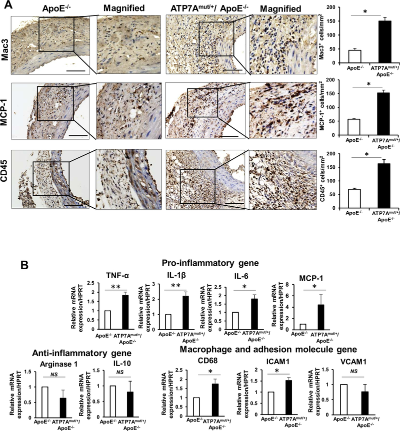
Figure 3. ATP7A dysfunction promotes proinflammatory responses in AAA.
(A and B) Abdominal aorta tissues from ApoE−/− and ATP7Amut/+/ApoE−/− mice following 4 weeks of Ang II infusion were examined. A, Representative images of immunohistochemical staining for macrophages (Mac3) and Monocytes chemoattractant protein-1 (MCP-1) and quantification. B, Dynamic expression profiles of inflammatory genes expression measured by qPCR (n=4). *p<0.05.

