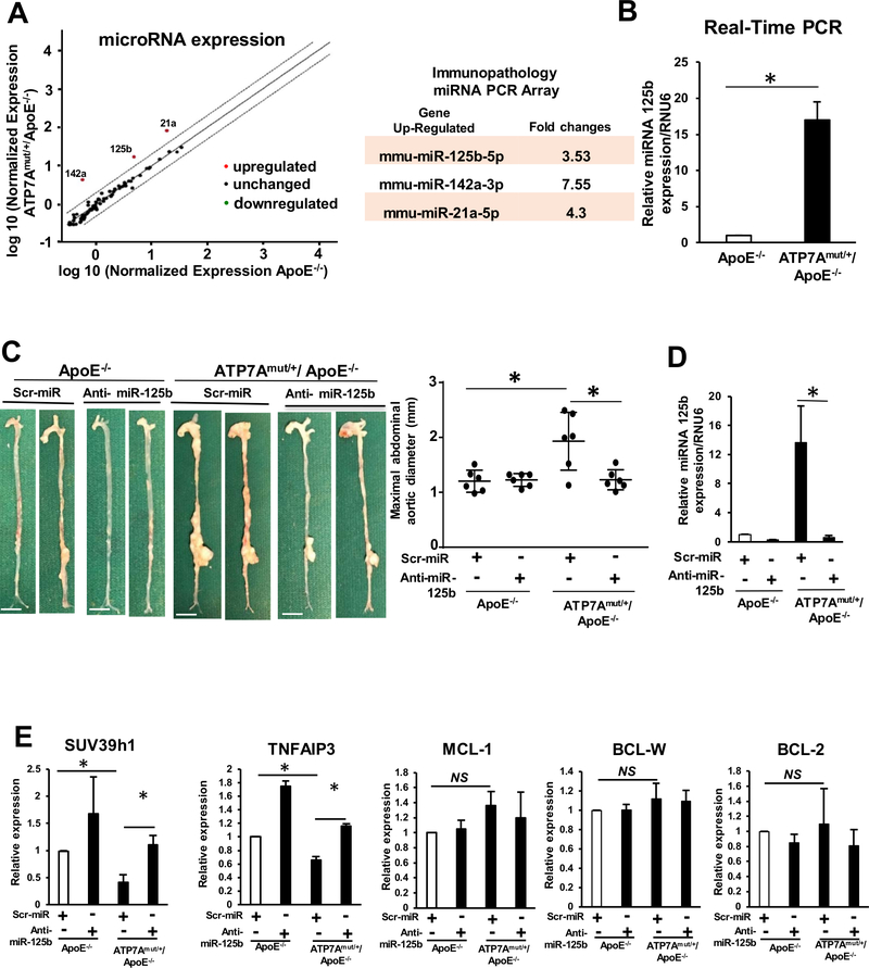
Figure 5. Anti-miRNA 125b treatment abolishes the development of Ang II-induced AA formation in ATP7Amut/+/ApoE−/− mice.
Abdominal aorta tissues from ApoE−/− and ATP7Amut/+/ApoE−/− mice following 4 weeks of saline or Ang II infusion were examined. (A) Identification of aortic miRNAs in ApoE−/− or ATP7Amut/+/ApoE−/− mice infused with Ang II for 4 weeks using the Qiagen miScript miRNA. Immunopathology array (n=3/group) and miScript PCR Array Data Analysis Tool. (B) miRNA 125b was validated using Real time-qPCR (n=4). (C,D,E) Representative images and maximum diameter of aorta (C), quantification of miRNA 125b expression (n=4) (D), andmiR-125b target gene expression analysis (n=4) using real-time qPCR (E) of mice with or without intravenous injection of LNA modified Anti-miRNA 125b or scrambled control. In C, scale bars: 3 mm. Maximal abdominal aortic diameter (Middle) with or without anti-miRNA 125b treatment (n=4). *p<0.05.

