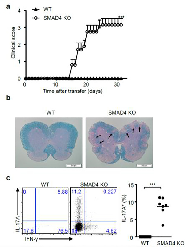
Figure 2. Adoptive transferred EAE model.
(a) CD4+ T cells isolated from wild-type 2D2 (WT) and Cd4-cre;Smad4fl/fl 2D2 (SMAD4 KO) mice were cultured with IL-21 and TGF-β receptor I inhibitor, and transferred into irradiated wild-type recipient mice on day0. Pertussis toxin was i.p. injected on day 0 and day 2. The EAE onset is day15. After 31 days, clinical scores (Mean ± SEM, n=10), (b) pathological and (c) flow cytometry and statistical analysis of EAE are shown. Lesions were indicated by the arrows in the figure.

