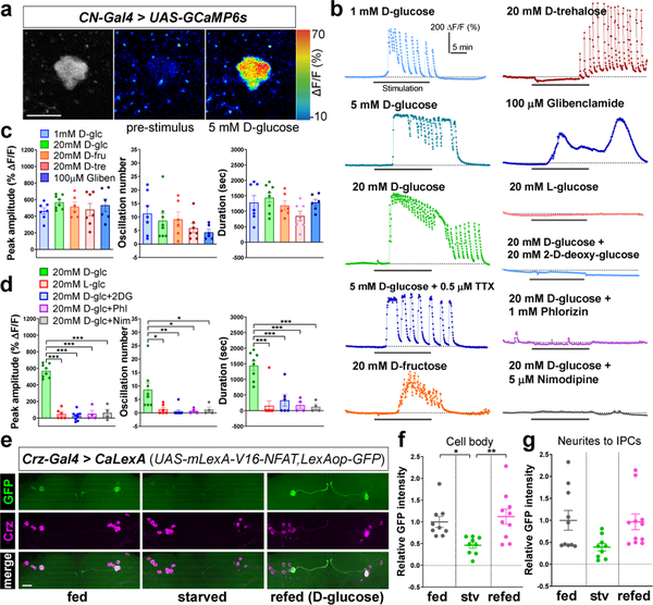
Figure 2.
CN neurons are activated by nutritive sugars, but not by nonnutritive sugars. a, CN neurons expressing GCaMP before (middle) and after (right) D-glucose application. Scale bar, 5 μm. b-d, Representative traces (b) and quantifications (c-d) of calcium responses of CN neurons to D-glucose (D-glc), D-fructose (D-fru), D-trehalose (D-tre), and glibenclamide (Gliben) (c) and L-glucose (L-glc), and D-glucose mixed with 2-D-deoxy-glucose (2DG), phlorizin (Phl), or nimodipine (Nim) (d). e-g, Representative images revealed by native CaLexA-driven GFP and anti-Crz staining (e), and quantifications (f-g) of GFP intensity from CN cell bodies (f) and neurites to IPCs (g) of fed, starved, or refed flies carrying Crz-Gal4 and CaLexA; see Methods. Scale bar, 20 μm. Z-stacked projections are shown. *P < 0.05, **P < 0.01, ***P < 0.001; one-way ANOVA with Tukey post hoc test. See Supplementary Table 1 for the sample sizes and statistical analyses.

