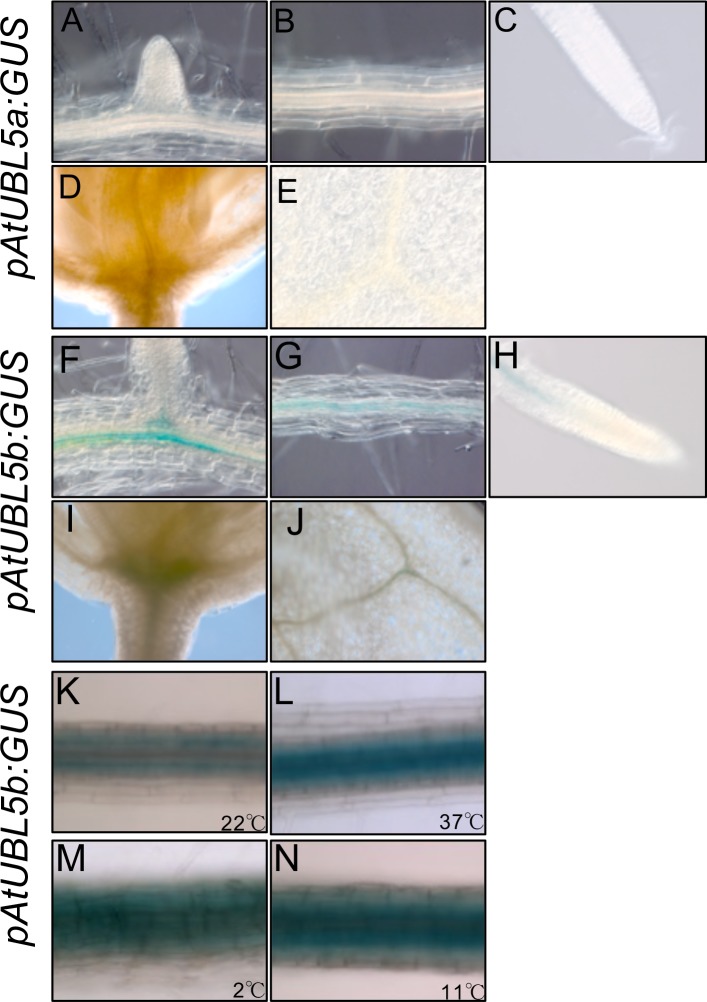
Fig 3. Histochemical analysis of AtUBL5a and AtUBL5b expression.
(A-E) GUS staining in pAtUBL5a:GUS plant tissue. (F-J) GUS activity in pAtUBL5b:GUS plant tissue. Emerging LR (lateral root) primordium (A and F), primary root (B and G), primary root apex (C and H), primordial vein (D and I), and leaf vein (E and J). (K-N) GUS staining in pAtUBL5b:GUS plants at 22°C, K, or under heat stress (37°C, L) and cold stress (2°C and 11°C, M and N).

