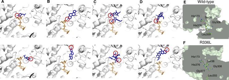
Fig 1. The complexes of UGT1A1 with coenzyme and substrates.
Pairs of UDPGA and substrates for wild-type UGT1A1 are as follows: A: UDPGA and AAP; B: UDPGA and E2; C: UDPGA and bilirubin; and D: UDPGA and SN-38. Top panels in each pair show the binding modes that can function in the conjugation reaction: the hydroxyl group of the substrate (red circle) is oriented toward UDPGA, which we call hydroxyl orientations; bottom panels show the binding modes that cannot function in the conjugation reaction. The UGT1A1 structures are shown as ribbon representations. UDPGA and substrates are shown as orange and blue sticks, respectively. (E and F) Cross-sectional images of wild-type UGT1A1 and R336L UGT1A1 structures highlighting the location of the glucuronidation and the amino acids of the coenzyme binding. The field of the conjugation process was disrupted in R336L.

