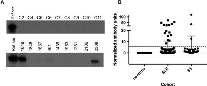
Figure 1.

Identification of patients with antibodies against retinoblastoma (Rb) protein. A, Sera from nine healthy control subjects and nine patients with systemic lupus erythematosus (SLE) (upper and lower panels, respectively) were used to immunoprecipitate 35S‐methionine–radiolabeled Rb generated by in vitro transcription/translation. Two of the patients with SLE shown in the panel had antibodies against Rb (1648 and 2308). The remaining seven patients with SLE were anti‐Rb negative, as were all nine of the controls. The leftmost lane of each panel (reference serum [ref ser]) denotes immunoprecipitations (IPs) performed with the positive reference serum used as a calibrator in each set of IPs. B, Levels of anti‐Rb antibody in the three cohorts tested. Antibodies were assayed as described in the Methods section. Each symbol denotes an individual serum sample; horizontal and vertical lines show the mean and SD. The upper broken line denotes the cutoff for assigning antibody positivity. SS, Sjögren syndrome.
