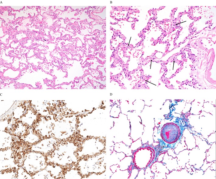
Figure 1.

Typical capillary proliferation (CP) and small‐vessel vasculopathy in lungs with advanced systemic sclerosis–related pulmonary fibrosis. (A) Diffuse distribution of CP (hematoxylin and eosin [H&E] stain; original magnification ×100); (B) Higher power example of CP; the alveolar walls have irregularly dilated capillaries with more than two layers (arrows) (H&E stain; original magnification ×200); (C) CP is demonstrated by highlighting endothelial cells, (CD31 immunohistochemistry stain; original magnification ×200); (D) Bronchovascular bundle demonstrating prominent pulmonary arterial intimal fibrosis (trichrome/elastin stain; original magnification ×100).
