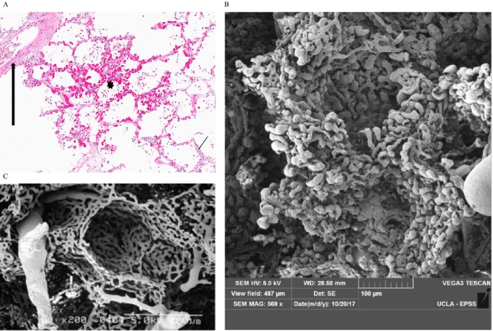
Figure 2.

Light microscopy evaluation of alveolar tissue from the systemic sclerosis–related pulmonary fibrosis–pulmonary hypertension (SSc‐PF‐PH) autopsy that demonstrates irregular alveolar wall capillary proliferation in areas without interstitial fibrosis: (A) capillary proliferation (asterisk) adjacent to normal artery (thick arrow) and normal alveolar capillaries (thin arrow) (hematoxylin and eosin [H&E] stain; original magnification ×200); (B) scanning electron microscopy (SEM) image of a vascular cast from the same SSc‐PF‐PH autopsy specimen highlighting extensive and crowded capillary duplication in the walls of two alveolar sacs. Note the abnormal pulmonary angiogenesis in the microvasculature with bulbous budding alternating with pinched areas in the alveolar capillary bed (SEM; original magnification ×569); (C) SEM of normal lung microvasculature and capillary bed. Note the much thinner capillary bed with more orderly arrangement of capillary architecture with few capillaries showing budding or abrupt termination of blind pouches (photos courtesy of Dr. Kazufumi Nakamura), (SEM, original magnification ×200.
