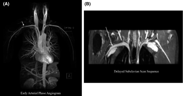
Figure 3.

Early arterial phase angiography versus venous phase imaging. A, Maximum intensity projection (MIP) reconstruction of an early arterial phase angiogram shows a pseudostenosis in the right subclavian artery in a healthy subject. B, A delayed phase dedicated subclavian scan shows that the suspected pseudostenosis seen during the early arterial phase is no longer visualized when imaging the same subject during the venous phase.
