. 2019 Nov 4;8:e51162. doi: 10.7554/eLife.51162

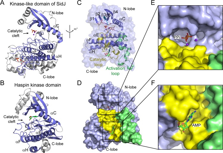
Figure 3. The core of SidJ adopts a protein kinase fold.

(A) Cartoon diagram of the kinase-like domain of SidJ. Secondary structure elements that are conserved in protein kinases are colored in blue. Ca2+ ions are shown as purple spheres while the pyrophosphate and AMP molecules are shown in sticks. (B) Cartoon representation of the kinase domain of Haspin kinase (PDB ID: 2WB6). The conserved structural core, colored in blue, is displayed with an orientation similar to that in panel (A). (C) An orthogonal view of the conserved secondary structural elements in the SidJ kinase-like domain. The N-lobe is comprised of five antiparallel β-strands and an αC helix. The C-lobe is primarily α helical. Secondary structural features are named according to PKA nomenclature. The activation loop is colored in green, the catalytic loop in yellow, and the glycine-rich loop in pink. Conserved residues within the kinase-like catalytic cleft are represented by sticks. (D) Surface representation of the SidJ kinase-like domain, depicting the catalytic cleft formed between the N- and C-lobes and the migrated nucleotide-binding site formed mainly by residues within the catalytic loop (yellow). The activation loop (green) makes close contact with the catalytic loop. (E) Enlarged view of the catalytic clefts outlined in (D). The kinase catalytic cleft contains two Ca2+ ions and a pyrophosphate (PPi) molecule. (F) Expanded view of the migrated nucleotide-binding pocket bound with an AMP.

Figure 3.

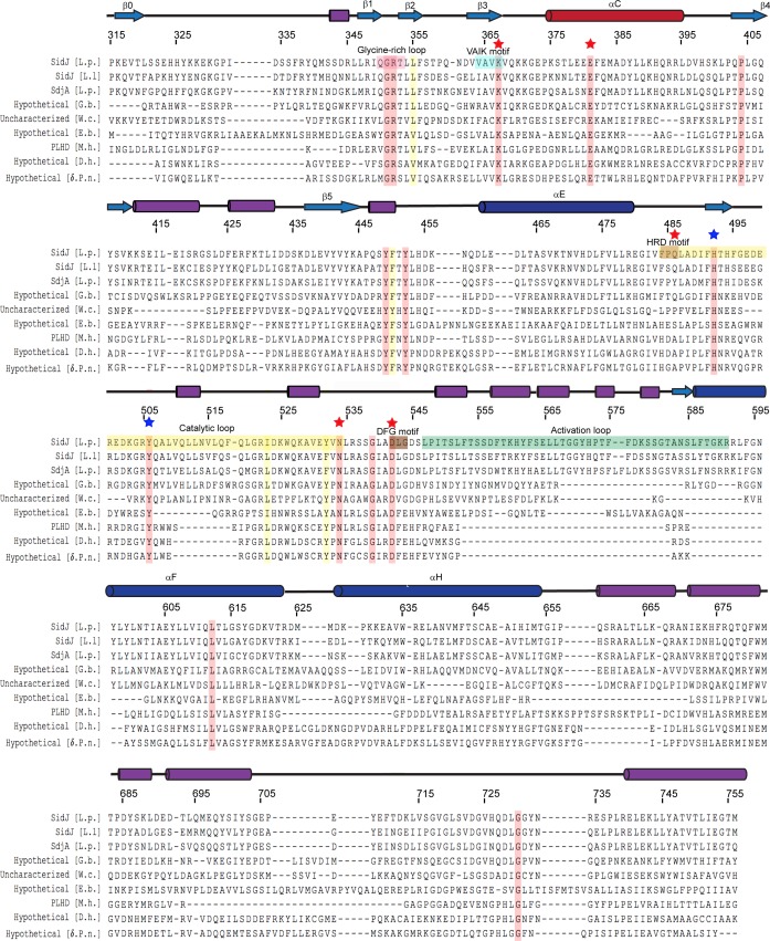
Figure 3—figure supplement 1. Multiple sequence alignment of SidJ kinase-like domain homologs.

Figure 3—figure supplement 1.

The NCBI BLAST server was used to identify proteins that are homologous to SidJ. Sequences corresponding to the kinase-like domain of SidJ (315–756) were aligned by Clustal Omega and colored using Multiple Align Show (http://www.bioinformatics.org/sms/index.html). SidJ residue numbers are marked above the alignment with secondary structural elements drawn above. Identical residues are highlighted in red and similar residues in yellow. Kinase catalytic residues located in the active site are marked with red stars, while residues at the migrated nucleotide-binding pocket are marked with blue stars. Conserved kinase motifs are highlighted as follows: glycine-rich loop (red), VAIK motif (blue), HRD motif (gold), catalytic loop (yellow), DFG motif (brown) and activation loop (green). NCBI accession numbers are as follows: SidJ, Legionella pneumophila, YP_096168.1; SidJ, Legionella longbeachae, WP_003634608.1; SdjA, Legionella pneumophila, YP_096515.1; hypothetical protein A3E83_09250, Gammaproteobacteria bacterium RIFCSPHIGHO2_12_Full_41_20, OGT46295.1; putative uncharacterized protein, Waddlia chondrophila 2032/99, CCB91008.1; hypothetical protein COB53_07685, Elusimicrobia bacterium, PCI37048.1; PBS lyase HEAT domain protein repeat-containing protein (PLHD), Methanosaeta harundinacea KUK97762.1; hypothetical protein, Desulfovibrio hydrothermalis WP_015335088.1; hypothetical protein, Delta proteobacterium NaphS2, WP_006420030.1.
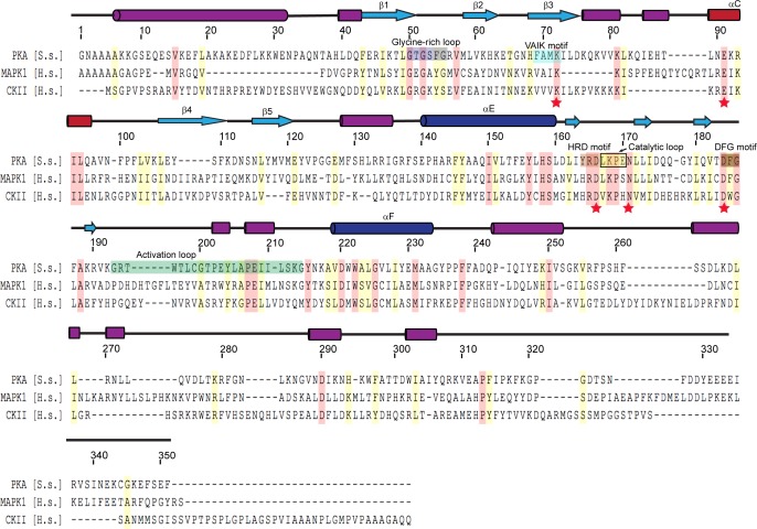
Figure 3—figure supplement 2. Multiple sequence alignment of representative protein kinases.

Figure 3—figure supplement 2.

The secondary structure of PKA is labeled above the sequence, and PKA residue numbers are marked on the top of the alignment. Identical residues are highlighted in red and similar residues in yellow. Kinase catalytic residues are marked with red stars and conserved kinase signature motifs are highlighted in a color scheme similar to that used for the SidJ kinase-like domain. Note that the catalytic loop in protein kinase contains only four amino acids, while the catalytic loop of SidJ is comprised of a large insertion (>40 amino acids, Figure 3—figure supplement 1). Accession numbers are as follows: cAMP-dependent protein kinase catalytic subunit alpha (PKA), Sus scrofa, P36887.4; mitogen-activated protein kinase (MAPK1), Homo sapiens, P28482; Casein kinase II subunit alpha (CKII), Homo sapiens, P68400.
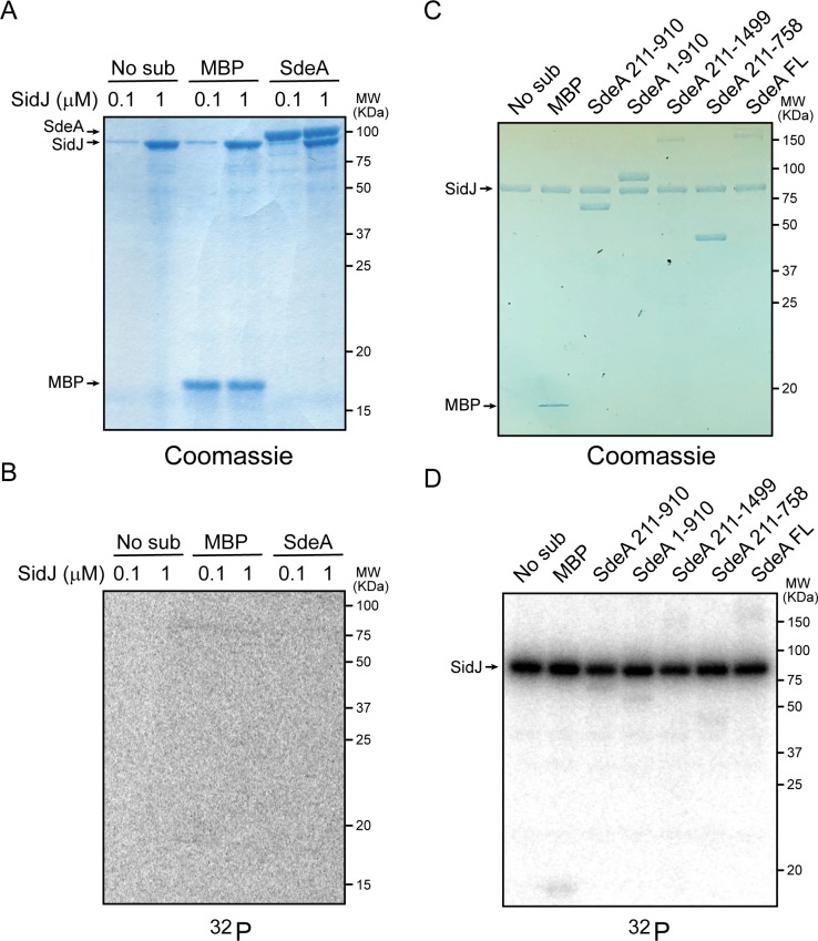
Figure 3—figure supplement 3. SidJ lacks canonical kinase activity but exhibits auto-AMPylation activity.

Figure 3—figure supplement 3.

(A) Two concentrations of SidJ (0.1 μM and 1 μM) were incubated with CaM, MgCl2 and [γ-32P]ATP without substrate, with MBP, and with SdeA 1–910 for 30 min at 37°C. Proteins were separated by 12% SDS-PAGE and visualized with Coomassie stain. (B) Autoradiogram of the gel shown in panel (A). Exposure time: 2 hr. (C) SidJ was incubated with CaM, MgCl2 and [α-32P]ATP without substrate, with MBP, and with the indicated recombinant SdeA truncations. Proteins were separated by 8% SDS-PAGE and visualized with Coomassie stain. (D) Autoradiogram of the gel shown in panel (C). Exposure time: 1 hr. The bands corresponding to SidJ showed strong radiographic signals, indicating auto-AMPylation of SidJ.